Talar Dome Lesion
What is Talar Dome Lesion?
Chondral Defects of Talas
A talar dome lesion is an injury to the cartilage and underlying bone of the talus within the ankle joint. It is also called an osteochondral defect (OCD) or osteochondral lesion of the talus (OLT). “Osteo” means bone and “chondral” refers to cartilage.
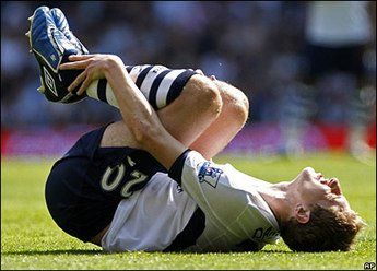
Talar dome lesions are usually caused by an injury, such as an ankle sprain. If the cartilage doesn’t heal properly following the injury, it softens and begins to break off. Sometimes a broken piece of the damaged cartilage and bone will “float” in the ankle.

Causes
Symptoms
The signs and symptoms of a talar dome lesion may include:
- Chronic pain deep in the ankle.
- An occasional clicking feeling.
- Ankle giving way.
- Swelling
Diagnosis
A talar dome lesion can be difficult to diagnose because the precise site of the pain can be hard to pinpoint. To diagnose this injury, the foot and ankle surgeon will question the patient about recent or previous injury and will examine the foot and ankle, moving the ankle joint to help determine if there is pain, clicking, or limitation of motion within that joint.
Sometimes the surgeon will inject the joint with an anaesthetic (pain-relieving medication) to see if the pain goes away for a while, indicating that the pain is coming from inside the joint.
X-rays are taken, and often an MRI or other advanced imaging tests are ordered to further evaluate the lesion and extent of the injury.
Non-surgical Treatment
Treatment depends on the severity of the talar dome lesion. If the lesion is stable (without loose pieces of cartilage or bone), one or more of the following non-surgical treatment options may be considered:
- Immobilization.
- Oral medications.
- Physical therapy.
When is Surgery Needed?
A talar dome lesion can be difficult to diagnose because the precise site of the pain can be hard to pinpoint. To diagnose this injury, the foot and ankle surgeon will question the patient about recent or previous injury and will examine the foot and ankle, moving the ankle joint to help determine if there is pain, clicking, or limitation of motion within that joint.
Sometimes the surgeon will inject the joint with an anaesthetic (pain-relieving medication) to see if the pain goes away for a while, indicating that the pain is coming from inside the joint.
X-rays are taken, and often an MRI or other advanced imaging tests are ordered to further evaluate the lesion and extent of the injury.
Complete and comprehensive spectrum of both diagnosis and treatments
QUICK LINKS
ABOUT
Specialist Foot and Ankle Surgery and Consultation in Manchester. Our specialist team of surgeons and podiatrists provide the highest level of expertise in the effective management of any foot and ankle condition. .
Manchester Foot And Ankle Clinic / North West OrthoSports is a Registered Trade Mark
North West OrthoSports / Manchester Foot & Ankle Clinic Ltd
93-107 . Lancefield Steet
Glasgow .G3 8HZ
Limited Company-387376
VAT Registration - 214 0705 54
Please See Our Legal Disclaimer on Online Medical Advice
All Rights Reserved | Manchester Foot And Ankle Clinic | Privacy Policy


