
Tarsal Coalition
What is Tarsal Coalition?
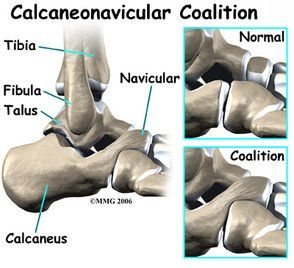
A tarsal coalition is an abnormal connection that develops between two bones in the back of the foot). This abnormal connection, which can be composed of bone, cartilage, or fibrous tissue, may lead to limited motion and pain in one or both feet.
Causes
Most often, tarsal coalition occurs during foetal development, resulting in the individual bones not forming properly. Less common causes of tarsal coalition include infection, arthritis, or a previous injury to the area.
Symptoms
While many people who have a tarsal coalition are born with this condition, the symptoms generally do not appear until ages nine to 16. Sometimes there are no symptoms during childhood.
The symptoms of tarsal coalition may include one or more of the following:
- Pain when walking or standing.
- Tired or fatigued legs.
- Muscle spasms in the leg, causing the foot to turn outward when walking.
- Flatfoot.
- Walking with a limp.
- Stiffness of the foot and ankle.
Diagnosis
A tarsal coalition is difficult to identify until a child’s bones begin to mature. Diagnosis includes obtaining information about the duration and development of the symptoms as well as a thorough examination of the foot and ankle. The findings of this examination will differ according to the severity and location of the coalition.
In addition to examining the foot, the surgeon will order x-rays. Advanced imaging studies may also be required to fully evaluate the condition.
Non-surgical Treatment
The goal of non-surgical treatment of tarsal coalition is to relieve the symptoms and reduce the motion at the affected joint. One or more of the following options may be used, depending on the severity of the condition and the response to treatment:
- Oral medications.
- Physical therapy.
- Steroid injections.
- Orthotic devices.
- Immobilization.
- Injection of an anaesthetic agent.
When is Surgery Needed?
A tarsal coalition is difficult to identify until a child’s bones begin to mature. Diagnosis includes obtaining information about the duration and development of the symptoms as well as a thorough examination of the foot and ankle. The findings of this examination will differ according to the severity and location of the coalition.
In addition to examining the foot, the surgeon will order x-rays. Advanced imaging studies may also be required to fully evaluate the condition.